animal cell electron microscope
20245936 No abstract available. Most cells are so tiny that they cannot be seen with the naked eye.

Animal Cell Tem Stock Image C025 2692 Science Photo Library
It uses a beam of electrons to illuminate the specimen.
. What microscope is used to view animal and plant. Browse 1553 animal cell microscope stock illustrations and vector graphics available royalty-free or start a new search to explore more great stock images and vector art. What can you see under a light.
The electron microscope is necessary to see smaller organelles like ribosomes macromolecular assemblies and macromolecules. Select from premium Animal Cell Microscope of the highest quality. Find Animal Cell Microscope stock photos and editorial news pictures from Getty Images.
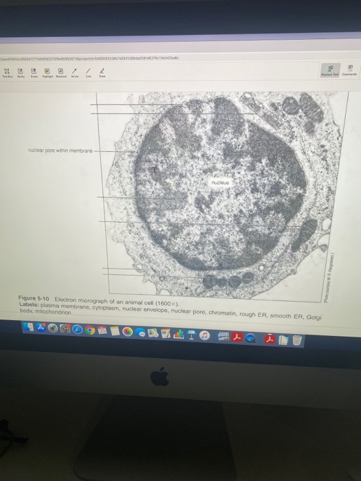
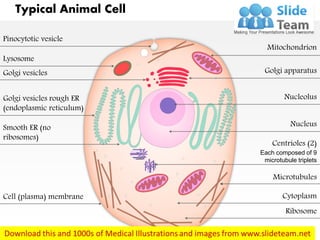
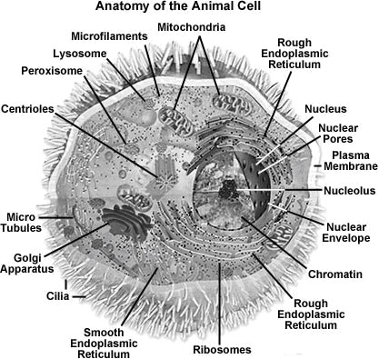
Below the basic structure is shown in the same animal cell on the left viewed with the light microscope and on the right with the transmission electron. The electron microscope is more powerful than the light microscope. Electron microscopy is in addition a valuable tool for the investigation of the most subtle effects of disease-causing organisms and toxic substances on animal cells and tissues.
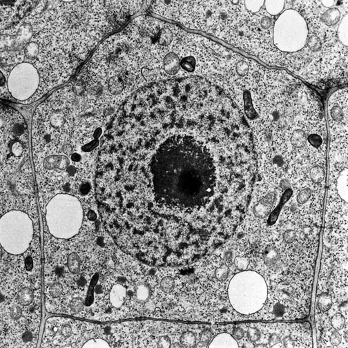
The Cell as Seen under the Electron Microscope. Animal cells have a basic structure. Up to 20 cash back You can use this royalty-free photo Electron microscope images of animal cells with nucleus and organelles for personal and commercial purposes according to.
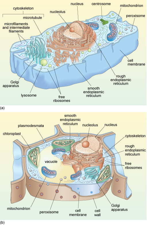
What microscope is used to view animal and plant. There are two types of electron microscopes. An animal cell also contains a cell membrane to keep all the organelles and cytoplasm contained but it lacks a cell wall.
Electron microscopy of animal cells Proc N Y State Assoc Public Health Lab. 2 days agoThanks to the advanced cryo-electron microscope in Umeå researchers have for the first time been able to take three-dimensional images of how the poliovirus forms and takes. Therefore scientists use microscopes to study cells.
A nm is 0001 uM. It was not until good light microscopes became available in. Electron microscopy of animal cells.
A cell is the smallest unit of life. An animal cell also contains a cell membrane to keep all the organelles and cytoplasm contained but it lacks a cell wall. I n a transmission electron microscope TEM the electrons pass through a very thin section of tissue much as light.
A typical animal cell is 1020 μm in diameter which is about one-fifth the size of the smallest particle visible to the naked eye.

File Anatomy And Physiology Of Animals Animal Cell Electron Microscope Jpg Wikimedia Commons
Cell Biology Wikipedia
A Tour Of The Cell View As Single Page
The Figure Below Is A Fine Structure Of A Generalized Animal Cell As Seen Under An Electron Tutorke
Electron Micrographs
What Does An Animal Cell Look Like Under An Electron Microscope Quora
Molecular Expressions Cell Biology Animal Cell Structure
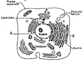
The Figure Below Is A Fine Structure Of A Generalized Animal Cell As Seen Under An Electron Microscope

Biology Notes For A Level 4 Cell Structure And Function
![]()
Electron Micrograph Animal Cell Hi Res Stock Photography And Images Alamy

Animal Cell Plant Cell Diagram Cell Diagram Animal Cell Structure
A Typical Animal Cell As Seen In An Electron Microscope Medical Ima
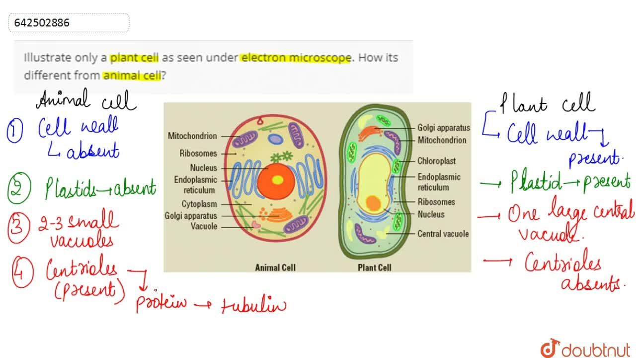
Illustrate Only A Plant Cell As Seen Under Electron Microscope How Its Different From Animal Cell

Amazon Com Eisco Plastic Animal Cell Electron Microscopic Structure 3d Model Toys Games

Animal Cell Definition Structure Parts Functions Labeled Diagram
Transmission Electron Microscopy Reveals Distinct Macrophage And Tick Cell Specific Morphological Stages Of Ehrlichia Chaffeensis Plos One

Ultrastructure
1 2 Ultrastructure Of Cells
Introduction To Cell